プラスミノーゲンアクチベーターインヒビター1
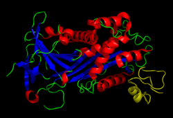
プラスミノーゲンアクチベーターインヒビター1またはプラスミノーゲン活性化抑制因子1(英: plasminogen activator inhibitor 1、略称: PAI-1)は、ヒトではSERPINE1遺伝子にコードされるタンパク質である。PAI-1の血中濃度の上昇は、血栓症やアテローム性動脈硬化のリスク因子となる[5]。
PAI-1はセリンプロテアーゼのインヒビター(セルピン)であり、プラスミノーゲン、そして線維素溶解(血栓の生理的分解)の活性化因子である組織プラスミノーゲンアクチベーター(tPA)やウロキナーゼ(uPA)に対する主要なインヒビターとして機能する。
他のPAIとしてはPAI-2があり、これは胎盤から分泌され、妊娠中にのみ多く存在する。さらに、プロテアーゼネキシンもtPAとuPAのインヒビターとして機能する。しかしながら、プラスミノーゲンアクチベーターの主要なインヒビターとなるのはPAI-1である。
遺伝子
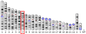
編集PAI-1をコードするSERPINE1遺伝子は7番染色体(7q21.3-q22)に位置する。プロモーター領域には4G/5Gと呼ばれる一般的多型がみられる。5Gアレルは4Gアレルよりもわずかに転写活性が低い[6]。
機能
編集PAI-1の主要な機能の1つが、プラスミノーゲンの切断によるプラスミン形成を担う酵素であるuPAの阻害である。プラスミンは自身も細胞外マトリックスを分解する機能を持ち、マトリックスメタロプロテアーゼとともに機能する[7]。PAI-1はuPAの活性部位に結合し、プラスミンの形成を防ぐ。PAI-1にはさらなる阻害機構も存在し、uPA/uPA受容体複合体に結合することで、uPA受容体の分解を引き起こす[8]。このように、PAI-1はtPAやuPAといったセリンプロテアーゼを阻害し、それによって血栓を分解する生理的過程である線維素溶解の阻害因子として機能する。さらにPAI-1はフーリンの活性阻害を介してマトリックスメタロプロテアーゼの活性化も阻害する[9]。
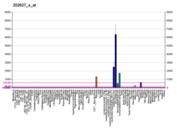
PAI-1は主に血管内皮細胞によって産生されるが、脂肪組織など他の組織からも分泌される[10]。
疾患における役割
編集PAI-1の先天性欠乏症が報告されており、線維素溶解が適切に抑制されないために出血傾向となる[11]。一方、線維症、がん、肥満、メタボリックシンドロームなどさまざまな疾患でPAI-1の上昇がみられる[12]。過剰なPAI-1はタンパク質分解活性を抑制することで細胞外マトリックスを安定化し、創傷治癒過程において線維形成を促進することで、組織の線維化に寄与する[13]。また、PAI-1は細胞老化を誘導する場合があり[14]、細胞老化随伴分泌現象(SASP)の構成要素となっている場合がある[15]。アンジオテンシンIIはPAI-1の合成を増加させ、アテローム性病変の形成を加速する[16]。
薬理
編集相互作用
編集出典
編集- ^ a b c GRCh38: Ensembl release 89: ENSG00000106366 - Ensembl, May 2017
- ^ a b c GRCm38: Ensembl release 89: ENSMUSG00000037411 - Ensembl, May 2017
- ^ Human PubMed Reference:
- ^ Mouse PubMed Reference:
- ^ “PAI-1 and atherothrombosis”. Journal of Thrombosis and Haemostasis 3 (8): 1879–1883. (August 2005). doi:10.1111/j.1538-7836.2005.01420.x. PMID 16102055.
- ^ Tziastoudi, Maria; Dardiotis, Efthimios; Pissas, Georgios; Filippidis, Georgios; Golfinopoulos, Spyridon; Siokas, Vasileios; Tachmitzi, Sophia V.; Eleftheriadis, Theodoros et al. (2021-11-25). “Serpin Family E Member 1 Tag Single-Nucleotide Polymorphisms in Patients with Diabetic Nephropathy: An Association Study and Meta-Analysis Using a Genetic Model-Free Approach”. Genes 12 (12): 1887. doi:10.3390/genes12121887. ISSN 2073-4425. PMC 8701119. PMID 34946835.
- ^ Lijnen, H. R. (2001-07). “Plasmin and matrix metalloproteinases in vascular remodeling”. Thrombosis and Haemostasis 86 (1): 324–333. ISSN 0340-6245. PMID 11487021.
- ^ “Obesity and breast cancer: the roles of peroxisome proliferator-activated receptor-γ and plasminogen activator inhibitor-1”. PPAR Research 2009: 345320. (2009). doi:10.1155/2009/345320. PMC 2723729. PMID 19672469.
- ^ Hohensinner, Philipp J.; Baumgartner, Johanna; Kral-Pointner, Julia B.; Uhrin, Pavel; Ebenbauer, Benjamin; Thaler, Barbara; Doberer, Konstantin; Stojkovic, Stefan et al. (2017-10). “PAI-1 (Plasminogen Activator Inhibitor-1) Expression Renders Alternatively Activated Human Macrophages Proteolytically Quiescent”. Arteriosclerosis, Thrombosis, and Vascular Biology 37 (10): 1913–1922. doi:10.1161/ATVBAHA.117.309383. ISSN 1524-4636. PMC 5627534. PMID 28818858.
- ^ Morange, P. E.; Alessi, M. C.; Verdier, M.; Casanova, D.; Magalon, G.; Juhan-Vague, I. (1999-05). “PAI-1 produced ex vivo by human adipose tissue is relevant to PAI-1 blood level”. Arteriosclerosis, Thrombosis, and Vascular Biology 19 (5): 1361–1365. doi:10.1161/01.atv.19.5.1361. ISSN 1079-5642. PMID 10323791.
- ^ Minowa, H.; Takahashi, Y.; Tanaka, T.; Naganuma, K.; Ida, S.; Maki, I.; Yoshioka, A. (1999). “Four cases of bleeding diathesis in children due to congenital plasminogen activator inhibitor-1 deficiency”. Haemostasis 29 (5): 286–291. doi:10.1159/000022514. ISSN 0301-0147. PMID 10754381.
- ^ Nam, Da-Eun; Seong, Hae Chang; Hahn, Young S. (2021). “Plasminogen Activator Inhibitor-1 and Oncogenesis in the Liver Disease”. Journal of Cellular Signaling 2 (3): 221–227. doi:10.33696/signaling.2.054. ISSN 2692-0638. PMC 8525887. PMID 34671766.
- ^ Ghosh, Asish K.; Vaughan, Douglas E. (2012-02). “PAI-1 in tissue fibrosis”. Journal of Cellular Physiology 227 (2): 493–507. doi:10.1002/jcp.22783. ISSN 1097-4652. PMC 3204398. PMID 21465481.
- ^ “Hepatic stellate cell senescence in liver fibrosis: Characteristics, mechanisms and perspectives”. Mechanisms of Ageing and Development 199: 111572. (October 2021). doi:10.1016/j.mad.2021.111572. PMID 34536446.
- ^ “Cellular senescence in the aging and diseased kidney”. Journal of Cell Communication and Signaling 12 (1): 69–82. (March 2018). doi:10.1007/s12079-017-0434-2. PMC 5842195. PMID 29260442.
- ^ van Leeuwen, R. T.; Kol, A.; Andreotti, F.; Kluft, C.; Maseri, A.; Sperti, G. (1994-07). “Angiotensin II increases plasminogen activator inhibitor type 1 and tissue-type plasminogen activator messenger RNA in cultured rat aortic smooth muscle cells”. Circulation 90 (1): 362–368. doi:10.1161/01.cir.90.1.362. ISSN 0009-7322. PMID 8026020.
- ^ “Tiplaxtinin, a novel, orally efficacious inhibitor of plasminogen activator inhibitor-1: design, synthesis, and preclinical characterization”. Journal of Medicinal Chemistry 47 (14): 3491–3494. (July 2004). doi:10.1021/jm049766q. PMID 15214776.
- ^ “Characterization of the Annonaceous acetogenin, annonacinone, a natural product inhibitor of plasminogen activator inhibitor-1”. Scientific Reports 6: 36462. (November 2016). Bibcode: 2016NatSR...636462P. doi:10.1038/srep36462. PMC 5120274. PMID 27876785.
- ^ “Plasminogen activator inhibitor-1 antagonist TM5441 attenuates Nω-nitro-L-arginine methyl ester-induced hypertension and vascular senescence”. Circulation 128 (21): 2318–2324. (November 2013). doi:10.1161/CIRCULATIONAHA.113.003192. PMC 3933362. PMID 24092817.
- ^ “Acute phase protein alpha 1-acid glycoprotein interacts with plasminogen activator inhibitor type 1 and stabilizes its inhibitory activity”. The Journal of Biological Chemistry 276 (38): 35305–35311. (September 2001). doi:10.1074/jbc.M104028200. PMID 11418606.
関連文献
編集- “[Type 1 plasminogen activator inhibitor: its role in biological reactions]”. [Rinsho Ketsueki] the Japanese Journal of Clinical Hematology 32 (5): 487–489. (May 1991). PMID 1870265.
- “Plasminogen activator inhibitor 1: physiological and pathophysiological roles”. News in Physiological Sciences 17 (2): 56–61. (April 2002). doi:10.1152/nips.01369.2001. PMID 11909993.
- “Plasminogen activator inhibitor-1 and the kidney”. American Journal of Physiology. Renal Physiology 283 (2): F209–F220. (August 2002). doi:10.1152/ajprenal.00032.2002. PMID 12110504.
- “Association between platelet activation and fibrinolysis in acute stroke patients”. Neuroscience Letters 384 (3): 305–309. (August 2005). doi:10.1016/j.neulet.2005.04.090. PMID 15916851.
- “Interaction of plasminogen activator inhibitor type-1 (PAI-1) with vitronectin (Vn): mapping the binding sites on PAI-1 and Vn”. Biological Chemistry 383 (7–8): 1143–1149. (2003). doi:10.1515/BC.2002.125. PMID 12437099.
- “The structural basis for the pathophysiological relevance of PAI-I in cardiovascular diseases and the development of potential PAI-I inhibitors”. Thrombosis and Haemostasis 91 (3): 425–437. (March 2004). doi:10.1160/TH03-12-0764. PMID 14983217.
- “Plasminogen activator inhibitor-I and tumour growth, invasion, and metastasis”. Thrombosis and Haemostasis 91 (3): 438–449. (March 2004). doi:10.1160/TH03-12-0784. PMID 14983218.
- “Urokinase-type plasminogen activator (uPA) and its inhibitor PAI-I: novel tumor-derived factors with a high prognostic and predictive impact in breast cancer”. Thrombosis and Haemostasis 91 (3): 450–456. (March 2004). doi:10.1160/TH03-12-0798. PMID 14983219.
- “Plasminogen activator inhibitor type 1: the two faces of the same coin”. Current Opinion in Nephrology and Hypertension 13 (1): 39–44. (January 2004). doi:10.1097/00041552-200401000-00006. PMID 15090858.
- “Plasminogen activator inhibitor-type 1: its plasma determinants and relation with cardiovascular risk”. Thrombosis and Haemostasis 91 (5): 861–872. (May 2004). doi:10.1160/TH03-08-0546. PMID 15116245.
- “Pleiotropic functions of plasminogen activator inhibitor-1”. Journal of Thrombosis and Haemostasis 3 (1): 35–45. (January 2005). doi:10.1111/j.1538-7836.2004.00827.x. PMID 15634264.
- “Plasminogen activator inhibitor-1: a common denominator in obesity, diabetes and cardiovascular disease”. Current Opinion in Pharmacology 5 (2): 149–154. (April 2005). doi:10.1016/j.coph.2005.01.007. PMID 15780823.
- “Historical analysis of PAI-1 from its discovery to its potential role in cell motility and disease”. Thrombosis and Haemostasis 93 (4): 631–640. (April 2005). doi:10.1160/TH05-01-0033. PMID 15841306.
- “[Role of PAI-1 in gynaecological malignancies]”. Zentralblatt für Gynakologie 127 (3): 125–131. (June 2005). doi:10.1055/s-2005-836407. PMID 15915389.
- “Plasminogen activator inhibitor type 1 gene polymorphism and sepsis”. Clinical Infectious Diseases 41 (Suppl 7): S453–S458. (November 2005). doi:10.1086/431996. PMID 16237647.
- “Plasminogen activator inhibitor-1, adipose tissue and insulin resistance”. Current Opinion in Lipidology 18 (3): 240–245. (June 2007). doi:10.1097/MOL.0b013e32814e6d29. PMID 17495595.
外部リンク
編集- ペプチダーゼとその阻害因子に関するMEROPSオンラインデータベース: I04.020
- Plasminogen Activator Inhibitor 1 - MeSH・アメリカ国立医学図書館・生命科学用語シソーラス
- Overview of all the structural information available in the PDB for UniProt: P05121 (Plasminogen activator inhibitor 1) at the PDBe-KB.
